
Diagnostic Imaging of the Head
- Non-Neuro structures
DATE: October 9–10, 2026
LOCATION: Warsaw, Klinika Teodorowscy
LECTURER: Prof. Tobias Schwarz
TIME: 9:00 AM – 5:00 PM
INTERPRETATION: English–Polish
THE TRAINING CONSISTS OF TWO PARTS:
» Theoretical introduction to diagnostic imaging
» Case studies at the workstations
WORKSHOP COORDINATOR: Dr. Piotr Teodorowski, DVM, Phd
FEES:
Early Bird: PLN 4400 net (until December 31, 2025)
Regular Price: PLN 5500 net (after December 31, 2025)
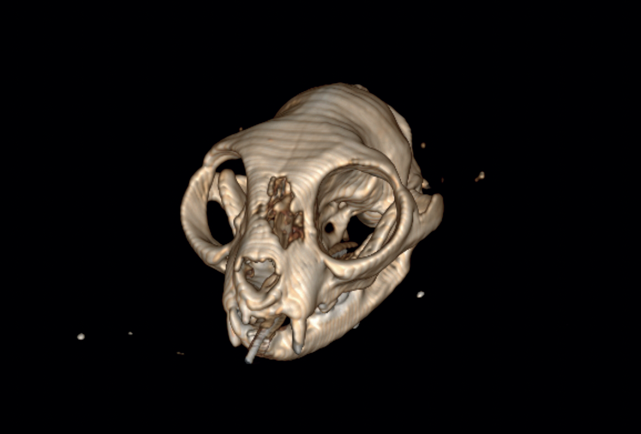
TARGET AUDIENCE: Veterinarians who wish to expand their knowledge of diagnostic imaging, gain a new perspective on radiography, and better understand in which cases it is advisable to extend diagnostics with CT and MRI.
*Limited number of participants
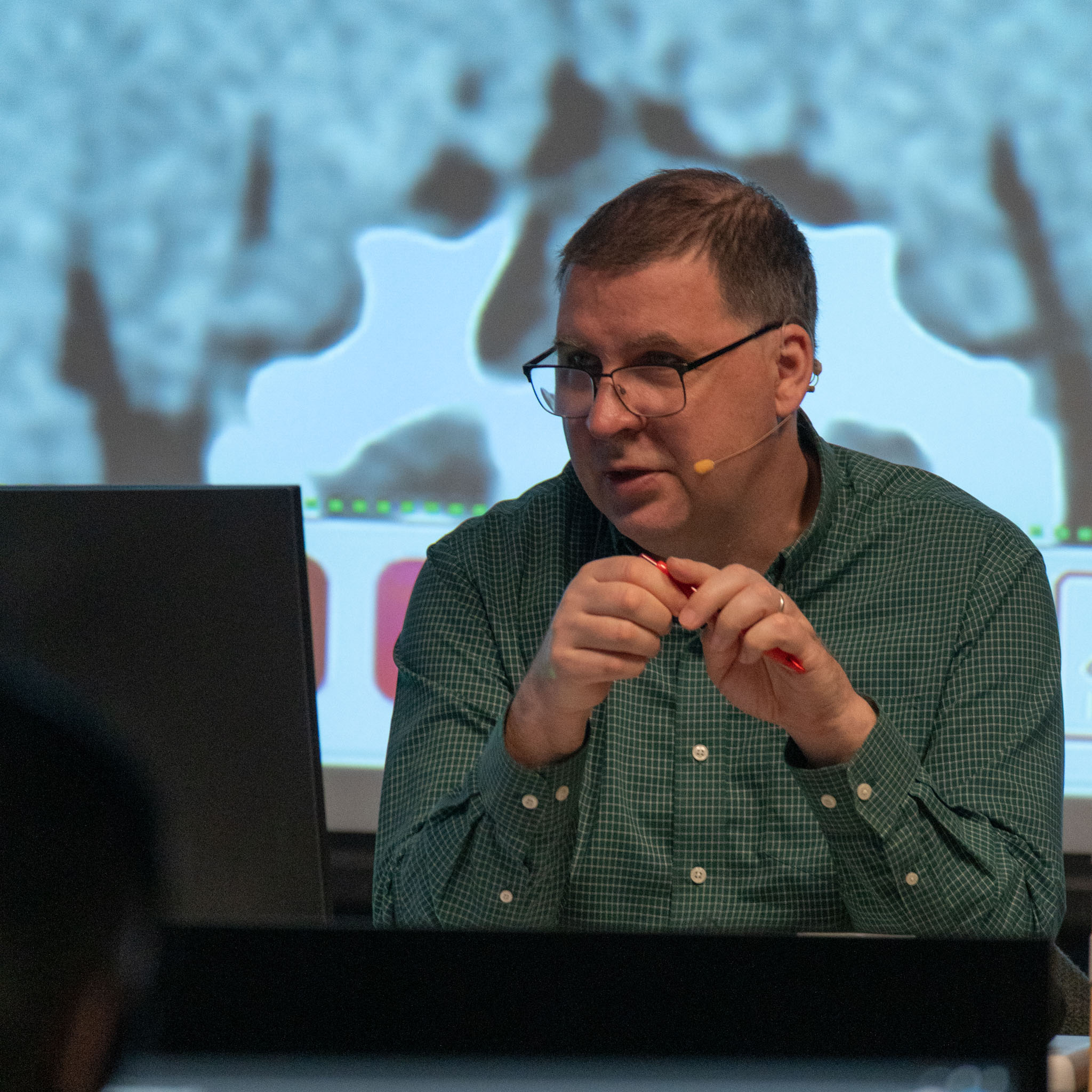
DVR, DipECVDI, DACVR, FRCVS Tobias Schwarz
Specialist in Veterinary Diagnostic Imaging
Graduated in veterinary medicine from Humboldt University and the Free University of Berlin, where he also completed a doctoral thesis on the application of computed tomography of the nasal cavity in dogs. At the same time, he earned a Master of Arts degree in Scandinavian Studies from the Free University of Berlin.
Following his studies, he undertook a residency in veterinary diagnostic imaging at the University of Glasgow Veterinary School (UK). He holds several prestigious international qualifications, including Diplomate of the American College of Veterinary Radiology (DipACVR), Diplomate of the European College of Veterinary Diagnostic Imaging (DipECVDI), the Diploma in Veterinary Radiology (DVR), and the Certificate in Veterinary Radiology (CertVR). He is also PennHIP certified.
His professional interests focus on modern imaging techniques, in particular the role of radiography and computed tomography in the diagnosis of diseases in dogs and cats.
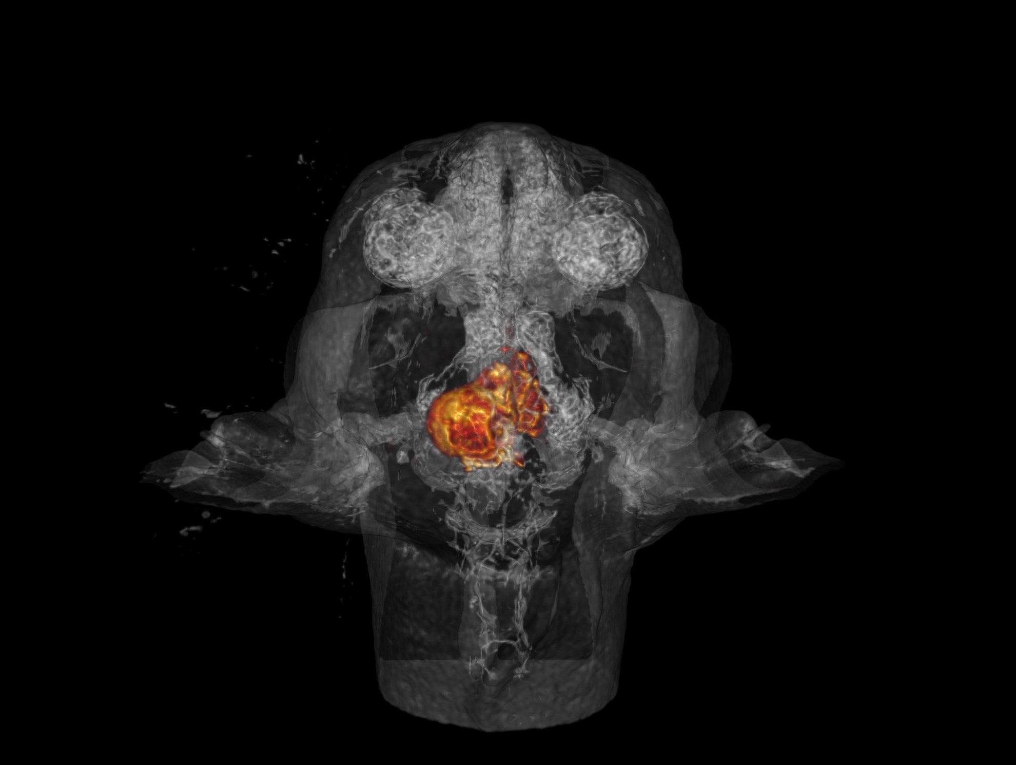
OUTLINE PROGRAM
Nasal CT
Dental & Masticatory CT
Oral Cavity, Salivary Gland & Tongue CT
CT of the Ear & Head Lymph Nodes
CT & MRI of the Eye and Orbital Cavity
CT of the Larynx, Pharynx & Soft Tissue Neck
Practical session: CT of the head – Non-Neuro structures (Parts 1–4)
DURING THE TRAINING YOU WILL
acquire a solid theoretical foundation in the imaging of head structures
learn how to interpret CT studies of the nasal cavity, useful in the diagnosis of neoplasia, inflammatory diseases, and foreign bodies
discover the applications of CT in veterinary dentistry for the evaluation of dentition, the masticatory apparatus, and oral pathology
understand how to effectively diagnose diseases of the salivary glands and tongue using computed tomography
gain insight into the role of CT and MRI in the diagnosis of ocular and orbital diseases, critical tools in challenging clinical cases
expand your knowledge of the imaging of the ear and head lymph nodes
learn how to perform and interpret CT examinations of the pharynx, larynx, and cervical soft tissues
gain valuable experience in image interpretation based on real clinical cases
benefit from up-to-date expert knowledge drawn from the lecturer’s extensive clinical and teaching experience
take advantage of the opportunity to exchange experiences with the instructor and fellow veterinarians
Sign up for the workshop:
Technology Partner:
GE HealthCare

Hosted by:
Klinika Teodorowscy Sp. z o.o.
7 Powstańców Street
43-180 Orzesze
WORKSHOP VENUE:
Klinika Teodorowscy
28 Maryli Street
02-842 Warszawa